


Pancreatic cancer
Orthotopic pancreatic cancer models support high-resolution monitoring of tumor progression, metastasis, and treatment response.
Breast cancer
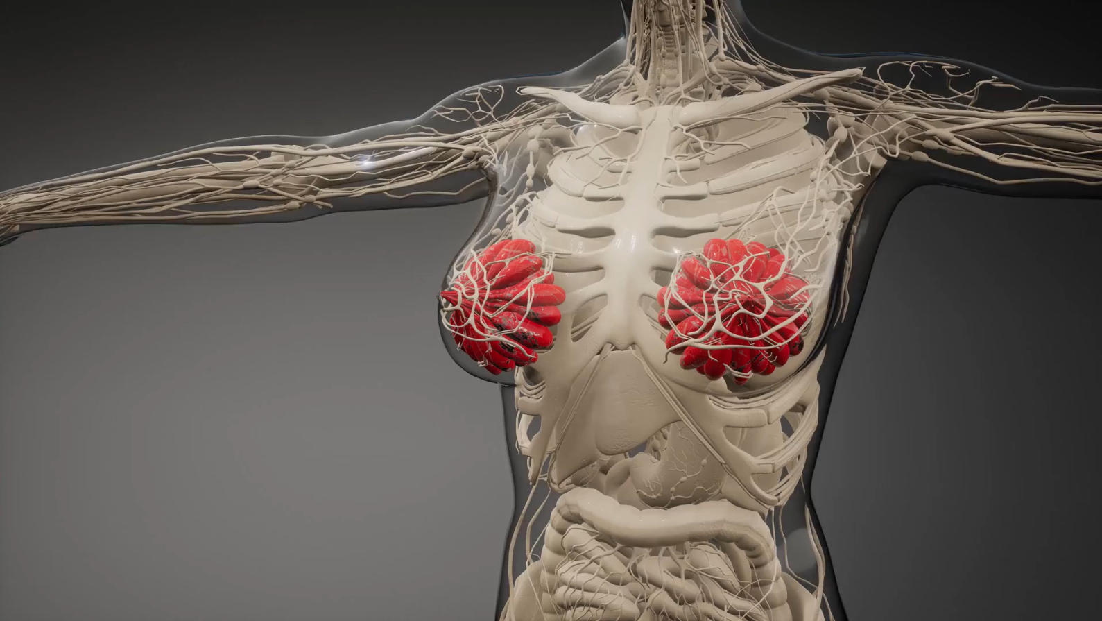
Our orthotopic breast cancer model provides a clinically relevant platform for studying primary tumor growth, metastatic spread, and therapeutic response.

Glioblastoma
Our orthotopic glioblastoma (GBM) model provides a clinically relevant platform for evaluating tumor growth, invasion, and therapeutic response.

Lung cancer
Our orthotopic lung cancer model provides a clinically relevant platform for studying tumor growth, metastatic spread, and therapeutic response.

Ovarian cancer
Ovarian cancer models include orthotopic and disseminated systems reflecting primary tumor growth and peritoneal metastasis.

Leukemia
Leukemia models recapitulate systemic disease and bone marrow interactions, enabling sensitive tracking of progression and treatment response.
Renal cell carcinoma
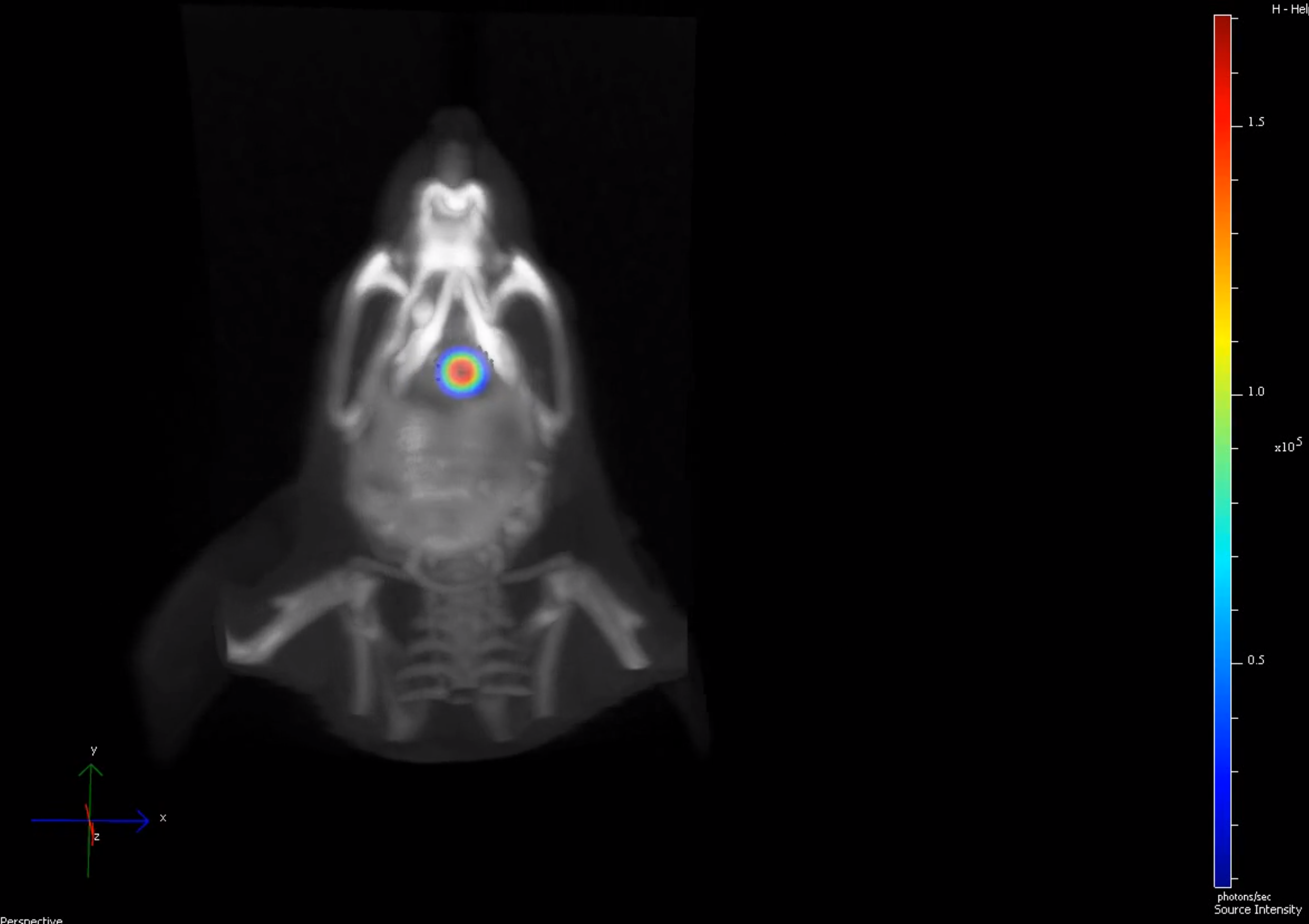
Renal cell carcinoma models reflect the vascularized and metastatic nature of kidney tumors.

Colorectal cancer
Colorectal cancer models replicate tumor growth in the colon and spread to distant organs such as the liver and lungs.