

Pancreatic cancer
Preclinical orthotopic pancreatic cancer models for in vivo studies of primary tumor growth, metastatic progression and therapeutic response.
Scientific overview
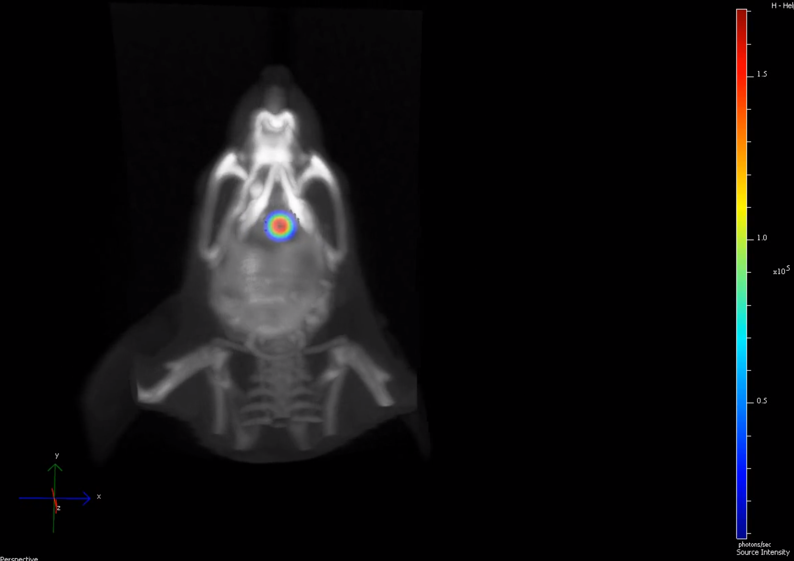
Our orthotopic pancreatic cancer model provides a clinically relevant platform for studying primary tumor growth, metastatic progression and therapeutic response in vivo. Longitudinal bioluminescence imaging (BLI), combined with 3D imaging and CT, enables quantitative monitoring and spatial localization of pancreatic tumor progression and metastatic spread.
Our orthotopic pancreatic cancer mouse model uses patient-derived or cell line-derived xenografts expressing luciferase, implanted directly into the pancreas to establish primary tumors in vivo. Longitudinal bioluminescence imaging (BLI) enables quantitative monitoring of primary tumor growth and metastatic progression in the same animal over time.
BLI can be combined with 3D imaging and CT to spatially localize pancreatic tumors and metastatic sites. Near-infrared fluorescent probes can also be incorporated to investigate tumor-associated processes including angiogenesis, apoptosis, metabolism and hypoxia. Studies can be complemented by blood sampling, histology and molecular analyses for further characterization of treatment effects.
Key capabilities
- Orthotopic pancreatic implantation
- PDX & cell-line xenograft models
- Longitudinal BLI + CT imaging
- Primary tumor & metastasis monitoring
Segment models
Explore our cancer models

Pancreatic cancer
Orthotopic pancreatic cancer models support high-resolution monitoring of tumor progression, metastasis, and treatment response.

Explore modell →

Breast cancer
Our orthotopic breast cancer model provides a clinically relevant platform for studying primary tumor growth, metastatic spread, and therapeutic response.

Explore modell →

Glioblastoma
Our orthotopic glioblastoma (GBM) model provides a clinically relevant platform for evaluating tumor growth, invasion, and therapeutic response.
Explore modell →

Lung cancer
Our orthotopic lung cancer model provides a clinically relevant platform for studying tumor growth, metastatic spread, and therapeutic response.

Explore modell →

Ovarian cancer
Ovarian cancer models include orthotopic and disseminated systems reflecting primary tumor growth and peritoneal metastasis.

Explore modell →

Leukemia
Leukemia models recapitulate systemic disease and bone marrow interactions, enabling sensitive tracking of progression and treatment response.
Explore modell →

Renal cell carcinoma
Renal cell carcinoma models reflect the vascularized and metastatic nature of kidney tumors.

Explore modell →

Colorectal cancer
Colorectal cancer models replicate tumor growth in the colon and spread to distant organs such as the liver and lungs.
Explore modell →
Let us to contribute
to your growth
Do you want to learn more about our work and how we can help you?